Articles
Articles Explained
Multi-slice CT Explained
Our new SOMATOM Sensation CT scanner allows the routine use of high performance Multi-slice CT, providing extended coverage of anatomical areas while producing images with higher detail:
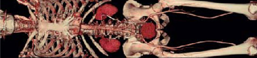
- Vasculature evaluation of the head, neck, thorax, abdomen, pelvis and extremities using CT Angiography.
- Evaluation of osteopathic and traumatic alterations of bone surface using 3-dimensional imaging.
- should be Evaluation of ischemic stroke injuries with the help of Perfusion CT, allowing physicians to meet the critical time window for effective treatment by providing results in less than 10 minutes.
- Computed tomography angiograms (CTA) excel at detecting significant narrowing of a blood vessel or artery that could require catheter-based intervention or surgery (such as bypassing).
Benefits
- Unlike other imaging methods, CT scanning offers detailed views of many types of tissue including the lungs, bones, soft tissues and blood vessels.
- CT scanning is painless, noninvasive and accurate. CT scans are also faster than many of the alternatives. It is cost-efficient enough to be useful for a wide range of clinical problems.
- CT scans can identify normal and abnormal structures, making it a useful tool to guide radiotherapy, needle biopsies and other minimally invasive procedures. Sometimes, CT images are so detailed they completely eliminate the need for invasive exploratory surgery and surgical biopsy.
CT Breakthrough: Multi-Slice CT Imaging

A state of the art imaging process gathers images in a single breath hold, virtually eliminating motion artifacts and providing superior diagnostic information.
Optimal Patient Comfort. Eliminates the closed-in feeling which patients sometimes associate with CT scanning.
Outstanding Diagnostic Information. By enabling us to perform Multi-slice CT on a routine basis.
Single Breath Hold Scans. We can now scan geriatric, pediatric and trauma patients in a single breath hold, which results in more accurate diagnosis while also reducing examination time and increasing patient comfort.
Multi-Channel RF Technology Explained
High quality RF coils and an optimized RF chain allow image acquisition with up to 8 channels and up to 16 CP coil elements. All array coils are iPAT compatible, for faster, higher differentiated and more clinically relevant images.
Integrated Panoramic Array (lPA) allows flexible combinations of up to 16 CP coil elements from up to 4 different coils. Patients and technologists will enjoy the easy handling and set-up time: the lower port of the CP Head Array Coil and the entire CP Spine Array Coil stay on the table for many exams.
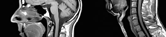
- MRI produces the clearest and most detailed images possible of soft-tissue structures of the body (e.g., the heart, lungs and liver). The high level of detail makes MRI an invaluable tool in early diagnosis and evaluation of tumors. It also reveals abnormalities that can be obscured by bone with other imaging methods.
- MRI can help physicians evaluate the function as well as the structure of many organs.
- MRI does not expose the patient to radiation. Also, the contrast material does not produce an allergic reaction. (We work with Berlex Laboratories, which manufactures the highest quality contrast materials available.)
- MRI offers a fast, noninvasive alternative to x-ray angiography for diagnosing problems ofthe heart and cardiovascular system.
- MR angiography (MRA) offers fast three-dimensional (3D) single breath-hold sequences for contrast enhanced MRA. As there is no radiation exposure, minimal invasivity and short examination time with high accuracy, MRA represents an alternative to digital arteriography.